

The size and fragility of infants make their surgeries among the most delicate and challenging to perform, especially pediatric surgeries to repair congenital heart defects such as hypoplastic left heart syndrome (HLHS). Whatever the nature of the critical congenital heart disease (critical CHD), a pediatric cardiac surgeon’s primary goal is to create or restore blood flow to a pathway so that oxygen-rich blood can reach the systemic circulation and a child can grow and thrive.
The American Heart Association estimates that 1% of children (~ 40,000) born each year in the United States have a heart defect that is generally discovered within the first 33 days of life.1 Most of these congenital defects are minor and may not cause problems until adulthood. However, within this 1%, one out of four babies (7,200) have critical CHD and require surgery within the first years of life. Symptoms in neonates with a heart defect include abnormal heart rhythms, blue-tinted skin, shortness of breath, failure to feed or develop normally, and swollen body tissue or organs.2
One critical CHD that requires surgery to save an infant’s life is hypoplastic left heart syndrome (HLHS). During pregnancy the left ventricle, mitral and aortic valves, and ascending aorta do not develop fully in the fetus, causing an underdeveloped left side of the heart which cannot pump oxygen-rich blood throughout the body when the child is born. 2
In addition to medical and nutritional treatments, a series of staged reconstructive surgeries are performed on HLHS babies to immediately increase blood flow to the body by bypassing the poorly functioning left ventricle and using the right ventricle as the heart’s primary heart pumping chamber. While not a cure, these initial three staged surgeries can save a neonate’s life.
The first surgery, a Norwood procedure, is usually performed within the first two weeks of a child’s life. During the surgery, the bottom of the pulmonary artery (which normally goes from the right ventricle to the lungs) is joined with the aorta. This new, bigger aorta exits from the right ventricle to the body to enable the neonate to get some oxygen-rich blood throughout their body. Then a shunt (Blalock–Thomas-Taussig Shunt) is created to move blood from (and through) the aorta to the lungs. Finally, a second shunt (Sano) is created to move blood from the right ventricle to the pulmonary artery and then to the lungs to become oxygenated.
The second or Bidirectional Glenn Shunt procedure is usually performed when an infant is 4 to 6 months of age. This procedure creates a direct connection between the pulmonary artery and the superior vena cava to reduce the right ventricle’s work by allowing blood returning from the upper part of the body via the superior vena cava to flow directly to the infant’s lungs.
The third and final staged reconstructive surgery is the Fontan procedure, performed when an infant is 18 months to three years old. During this surgery, surgeons connect the pulmonary artery to the inferior vena cava. This allows blood from the lower part of the body to go directly into the lungs. When oxygen-rich and oxygen-poor blood no longer mix in the heart, a child’s skin will no longer appear bluish.
Shortly after the Transonic Flowmeter was approved for human use in 1988, legendary pediatric cardiac surgeon Dr. Constantine Mavroudis, then at Children’s Hospital in Chicago, applied a Transonic Flowprobe to the aorta to measure cardiac output directly in mL/min during pediatric heart surgeries.3 He routinely measured ascending aorta flow before and after a corrective surgery. His patients ranged from 18 months to 12 years of age, with the usual age between 3-4 years.
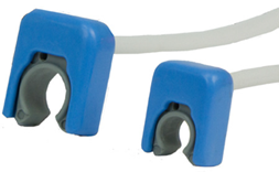
4-and 6-mm COnfidence Flowprobes
To accommodate the turbulent flows in the ascending aorta and improve accuracy, Transonic then developed a specialized four-crystal COnfidence® Flowprobe that is ideally suited to monitor cardiac output during surgical repair. Its slim, ergonomic profile creates a minimal footprint that allows the Flowprobe to fit in tight anatomical sites such as around the ascending aorta. A soft, pliable Flowprobe liner cushions and protects the vessel.
COnfidence Flowprobes continue to be valuable tools used by discriminating pediatric cardiac surgeons during complex reconstructive surgeries to repair critical CHDs in infants. The volume flow measurement data provided by the Flowprobes either confirm a surgeon’s clinical impressions that impact decision-making during the surgery or alert the surgeon to a potential problem at a time when it can be most readily addressed. 4
References:
- https://www.heart.org/en/health-topics/congenital-heart-defects/about-congenital-heart-defects
- Mai CT, Isenburg JL, Canfield MA, et al. for the National Birth Defects Prevention Network. National population-based estimates for major birth defects, 2010-2014. Birth Defects Res 2019; 1– 16. https://doi.org/10.1002/bdr2.1589. Last Reviewed:February 2, 2023. Source: National Center on Birth Defects and Developmental Disabilities, Centers for Disease Control and Prevention
- Mavroudis C, Backer CL, Kohr LM, Deal BJ, Stinios J, Muster AJ, Wax DF. Bidirectional Glenn shunt in association with congenital heart repairs: the 1(1/2) ventricular repair. Ann Thorac Surg. 1999 Sep;68(3):976-81; discussion 982.
- Kotani Y, Honjo O, Shani K, Merklinger SL, Caldarone C, Van Arsdell G. Is indexed preoperative superior vena cava blood flow a risk factor in patients undergoing bidirectional cavopulmonary shunt? Ann Thorac Surg. 2012 Nov;94(5):1578-83..