

 In science, pathophysiological mechanisms are unraveled by extensive, complex, and complementary combinations of in vitro and in vivo studies. This blog, the first in a series of three, will take you through one publication as an example of how to design an experimental approach to answer your research question.
In general, in vitro studies are faster, relatively easier to quantify results, typically less expensive, and focused on specific steps, elements and/or interactions of any given multilayered system. With protein knock-out and protein over-expressing cell lines for instance, it is possible to prove causal relationships between proteins and the processes in which they are involved.
In vivo studies are exponentially more complex models, as they encompass the whole body and therefore, all the mechanistic layers associated with physiological responses. For this reason, it is virtually impossible to completely isolate a given variable in vivo.
Simply put, experiments are designed to evaluate the role of “A” on “B”, and one must carefully strategize how to:
- Properly identify the baseline or "normal" levels for “A”. “A” can, for instance, be the serum level of a peptide, tissue protein content for a given receptor expression, blood pressure or heart rate levels under “control” conditions.
- Establish a way to increase or decrease “A’s” participation on the mechanism being studied. Enzyme substrates and receptor blockers are popular examples of pharmacological maneuvers used to challenge “A’s” contribution systemically. Tissue specific inducible knock-out and over-expressor models would be genetic approaches to influence the levels of “A”.
- Properly measure “B’s” response to “A’s” variation. In some cases, the parameter “B” can be easier to access (i.e. blood pressure measurements). In other stances, the specific variable “B” might be difficult to be evaluated under the experimental conditions (i.e. in conscious animals) and must be inferred from surrogate secondary variables. For example, stroke volume and/or ejection fraction are frequently used as indicatives of cardiac inotropy rather than using its direct measurements.
The intricacies involved in designing an in vivo experimental protocol can exponentially increase with the complexity of the mechanism to be studied, with its buffering, redundancy subsystems and their secondary effects. Sometimes these synergistic and/or oppositional effects can drastically affect variable “B” and lead to confounding results regarding the role of “A” on “B”. The following study to be discussed is a good example on how to identify these issues and how to address them through experimental design and careful data analysis.
In the “Role of endothelial nitric oxide in control of peripheral vascular conductance during muscle metaboreflex activation”, the study rationale is based on the following background:
- Muscle metaboreflex activation leads to an increase in blood pressure.
- This increase in blood pressure is due to an increase in Cardiac Output (CO) and not systemic vasoconstriction.
- Indeed, systemic vasodynamic activity leans towards epinephrine/vascular β2-receptors mediated vasodilation.
- The increase in CO represents an increase in blood flow and, therefore, increases sheer stress. Sheer stress is the primary stimulus for nitric oxide (NO) production by the endothelial cells.
- Although the systemic vasodilation is mostly mediated by epinephrine/vascular β2-receptors, it is possible that NO also contributes to the vasodilation.
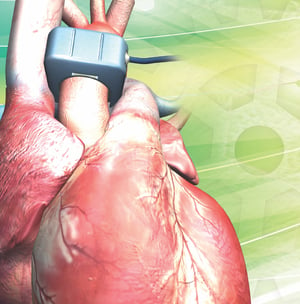
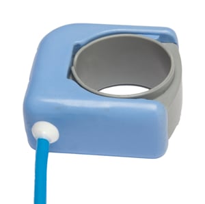 In the methodology section, the study describes the strategic instrumentation of mongrel canines that was used to independently evaluate the vasoactivity of systemic and regional blood flows. A transit-time ultrasound flow probe placed on the ascending aorta provided Cardiac Output (CO). A flow probe placed on the abdominal aorta provided direct information regarding hindlimbs blood flow, which are directly related to the muscle metaboreflex activation via ischemia induced by gradual occlusion of the abdominal aorta (by occluders). Blood pressure data (measured with a pressure catheter), combined with blood flow data, provides a vasoactivity index expressed, in this case, as conductance (calculated as blood flow divided by blood pressure). Once the muscle metaboreflex is activated by partial inflation of terminal aortic occluders during moderate treadmill exercise, the hindlimb blood flow is characterized as ischemic vasculature. The assessment of the non-ischemic vasculature is then calculated as CO (total blood flow) minus the abdominal aorta blood flow (ischemic vasculature). Again, the availability of blood pressure data allows for vascular conductance calculation for the non-ischemic vasculature.
Usually, the contribution of one variable/factor is assessed by its suppression rather than by its stimulation. The study evaluated muscle metaboreflex activation under normal conditions and under nitric oxide synthesis blockade by L-nitroarginine methyl ester (L-name). However, L-name administration affected other cardiovascular variables and is a clear example of how the manipulation of one factor has a chain effect down through the mechanistic interactions involved in the cardiovascular system modulation:
- Since NO is intrinsic to the vascular tonus, blood pressure (BP) increased by vasoconstriction.
- The increase in BP caused a baroreflex mediated decrease in Heart Rate (HR).
- The decrease in HR likely attenuated the CO response (increase) to muscle metaboreflex activation.
Administration of L-name affected both variables used to investigate the relationship between sheer stress (CO) and vasodilation (NO effect). Therefore, the interpretation of results as absolute values or as simple variation (delta) could lead to false conclusions. Since wall sheer stress denotes the primary stimulus for endothelial release of NO, the decrease in the CO response to exercise under L-name could affect the result’s interpretation due to an attenuation of the NO synthesis stimulus itself. Because NO blockade affects both basal hemodynamic values as well as responses to exercise, the data was analyzed as the relationship between CO (sheer stress stimulus) and vasodilation (NO effect). The slope of the linear regression calculated for these two variables revealed that the change in vascular tone (vasodilation) per unit change in flow (sheer stress) was not affected by L-name.
This study serves as a good example of how strategic, well-planned in vivo instrumentation and data analysis can lead to important advancements in research. Transonic has partnered with countless labs over our 37-year history – providing them with blood flow technologies that help to facilitate a wide array of study designs and requirements, including the study mentioned above. Stay tuned for part 2 in this series where we will discuss “Muscle metaboreflex-induced central blood volume mobilization in heart failure”