

 This second installment in our “Experimental Design Puzzles” blog series illustrates how the intricacies involved in designing an in vivo experimental protocol can exponentially increase due to the complexity of the mechanism to be studied and a variety of experimental limitations. In “Experimental Design Puzzles Part 1” we used the study “Role of endothelial nitric oxide in control of peripheral vascular conductance during muscle metaboreflex activation” to exemplify a careful and resourceful experimental design aimed to isolate effects within a complex multilayered regulatory mechanism in vivo.
Here we will discuss a second publication by the same group: “Muscle metaboreflex-induced central blood volume mobilization in heart failure”, which expands on the methodology and data analysis procedure mentioned on the first blog of these series.
For a better understanding of some common aspects of the methodology applied in each study, we would suggest reading our previous blog, and accessing the full paper: “Muscle metaboreflex-induced central blood volume mobilization in heart failure”, especially for the figures/graphs as they are referred to in this blog.
In the “Muscle metaboreflex-induced central blood volume mobilization in heart failure,” the study rationale is based on the following:
- Muscle metaboreflex activation leads to sustained increases in Cardiac Output (CO), Heart Rate (HR) and ventricular contractility.
- Increased (HR) and ventricular contractility alone are ineffective in sustaining the observed increases in (CO).
- This is due to the inverse relationship between cardiac output and ventricular filling pressure: “as cardiac output rises there is a translocation of blood volume from the central venous side to the peripheral arterial side of the circulation, and cardiac filling pressure falls.”
- During muscle metaboreflex activation, central venous blood volume mobilization can maintain ventricular filling thereby sustaining the increase in cardiac output triggered by the reflex.
-
- A previous study conducted by the same group explored this relationship between central venous blood volume mobilization and CO. Once CO was held constant, central venous pressure increased, acting as an index of central blood volume mobilization.
- In heart failure (HF), the muscle metaboreflex induced increases in CO and ventricular contractility are compromised.
- These impaired reflex responses can be explained by the characteristic HF ventricular dysfunction; however, it is unknown if the central blood volume mobilization mechanism is also impaired in systolic HF.
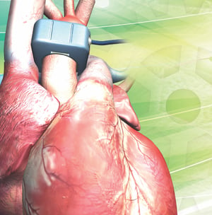
 Methods: As in the first blog of this series, a simplified version of the methodology is described, including the strategic instrumentation of mongrel canines that was used to independently evaluate the vasoactivity of systemic and regional blood flows, as well as the arterial and venous pressures. Three distinct surgeries were undertaken to achieve the complete and complex instrumentation:
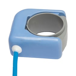
- Thoracic surgery - CO and pacing leads: A transit-time ultrasound flow probe placed on the ascending aorta to provide CO, and ventricular pacing electrodes were sutured to the left ventricular apex for future ventricular pacing (for HF induction and constant HR experiments).
- Retroperitoneal surgery - hindlimb blood flow, occluders and mean arterial pressure: A flow probe and occluders were placed around the abdominal aorta to provide direct information regarding hindlimb blood flow and activation of the muscle metaboreflex via induced ischemia by gradual occlusion of the occluders. A pressure catheter was placed in the abdominal aorta through a lumbar artery for arterial pressure measurement.
- Anterior cervical surgery - central venous pressure: A second catheter was placed in the arterial-caval junction through the jugular vein for central venous pressure (CVP) measurement.
All instrumentation was exteriorized through the scapular area.
Experiments: The following experiments were performed to activate muscle metaboreflex under different conditions; before and after induction of HF.
- Experiment 1 - Mild exercise with normal cardiac function: regular muscle metaboreflex activation experiments.
- Experiment 2 - Mild exercise with constant HR: reflex activation experiments conducted while the animals were connected to a fixed-rate pacemaker for HR to be held constant.
- Experiment 3 - Mild exercise with constant HR plus limited contractility. Reflex activation experiments conducted while the animals were connected to a fixed-rate pacemaker for HR to be held constant, and under atenolol treatment to limit ventricular contractility.
Data Analysis: All the variables evaluated in the study can be observed in Figure #1 and table #1 of the original paper, providing a summary of all data entries. Each reflex activation experiment has three distinct time points (rest, exercise, and reflex activation); these experiments were repeated under three different conditions (experiments #1, #2 and #3, all described above) before and after HF; resulting in a total of 18 data points for each hemodynamic variable.
Results and discussion: Note that the CO response is impaired in HF due to ventricular dysfunction, and the aim of the study was to determine if the central venous blood volume mobilization mechanism is also impaired by HF and could contribute to the impaired CO response. However, how can a variable be properly isolated in a condition riddled with so many variables and interactions? The study answers this methodological question by focusing on the relationship between CVP (index of central venous blood volume mobilization) and CO (directly dependent of CVP).
Figure #2 concentrates on this relationship between CVP and CO regarding only muscle metaboreflex activation. The best way to look at the trend depicted in that graph is to focus on the similarities in the lines connecting exercise and muscle metaboreflex activation (MMA), between control and HF data under the same experiments (1, 2 and 3). The best example can be observed in the data of experiment # 3 (constant HR plus limited contractility, represented with “square” symbols), comparing the line (between exercise and MMA) in control conditions (line between white square without cross and white square with cross) with HF (line between black square without cross and white square with cross). Although CO is higher and CVP lower under control conditions, the slopes of the lines connecting the same color symbols are remarkably similar.
In figure #3, the study further extends the analysis of these slopes, plotting the slopes data only and subjects the data to another linear regression between the three experimental settings in control and HF conditions. The combined data suggest that, despite all the variability between all the data points and hemodynamic measurements, central blood volume mobilization does not contribute to the attenuation of the reflex CO response in HF. This appears to be primarily due to poor ventricular performance.
This study is another excellent example of how strategic, well-planned in vivo instrumentation and data analysis can lead to important advancements in research. We hope this blog series will highlight some intricate experimental maneuvers used in the referred literature and, in some way assist researchers in developing their experimental designs. Transonic has participated in outstanding research over our 37-year history – providing labs with blood flow technologies and learning from their experiences and publications. Stay tuned for part three, the last in this series, where we will discuss conductance vs. resistance.